Dissecting Organs from the Deli
Fresh offal can provide cheap specimens of real internal organs to study.
Nearly every part of a mammalian body is edible. Some parts are tough and not pleasant to chew, but can still be eaten and contain nutritional value. Beef stomachs are available as “tripe”, intestines are often used as sausage casings, brains are a delicacy in some countries, and even bones can be boiled to make broth, or ground up into “bone meal”, which was once used as a dietary supplement and in animal feed. Only teeth, fur, and claws are useless as food.
The most tender and popular parts are usually the muscles or meat, and these are available everywhere. The internal organs are not as popular in American cuisine, but I can find sliced calf liver in most supermarkets, and whole organs of several kinds can often be found at ethnic or specialty meat markets. (Internal organs sold as food are called offal.) If you live in a city, you will probably not have much problem finding livers, hearts, kidneys, and tongues of sheep, pigs, and cows for sale. Sometimes you can find “sweetbreads,” which are various glands of cows or sheep. The only exceptions are brains and lungs. For some reason, lungs are illegal to sell as food in the United States, although they are popular in the cuisine of some other countries. As nearly as I can tell, brains are legal as food in the United States, but they are very rarely sold. The risks of brain-borne diseases and the difficulty in extracting them from the cranium probably don't help their popularity. I found frozen cow brains once in a specialty meat market, and now I really wish I had bought one, because I have never found them since.
Being able to buy fresh offal means that science students have a cheap and easy way to observe first-hand the structure and texture of real internal organs. Not only are these fresh organs cheaper than preserved organs from a science supply company, they are fresher, more realistic, and easier to work with. I purchased all of the following sheep organs from a nearby Middle Eastern deli for roughly two dollars apiece. I purchased the chicken organs and beef tripe from my corner grocery store. (Most of these organs are also available from cows, but cow organs are often too large to be convenient for dissecting. Middle Eastern markets don't normally carry pig organs, but you can often find them in Asian markets.) For the most part, kitchen knives and kitchen shears work fine for dissecting these organs. However, you may find a scalpel (or a razor-sharp hobby knife) to be helpful — much of the fabric in the kidney and the heart is very tough and difficult to cut with a kitchen knife.
Kidneys
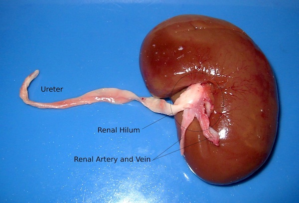
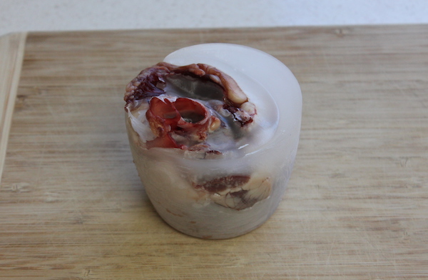
A kidney is a bean-shaped organ with one “doorway” on the concave side through which three different tubes come and go. This doorway into the kidney is officially named the hilum. (Lungs also have hila.) The three tubes are the ureter, which connects the kidney to the bladder, and two blood vessels which connect the kidney to the two major blood vessels alongside the spine. The two blood vessels are the renal artery, which connects to the aorta, and the renal vein, which connects to the vena cava. A clue to distinguishing between the renal artery and the renal vein is that arteries have muscular walls, and so are thicker and more elastic than veins.
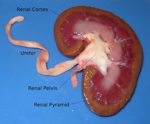
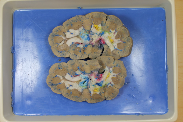
If you slice through a kidney along the midplane, you discover a complex interwoven structure inside. You might even think it's pretty. The three tubes branch out into three overlapping “tube trees,” and all three blend together in the spongy material around the edges of the organ — the cortex of the kidney. Between the hilum and the cortex, the tube systems pass back and forth through various branches and the distinctive renal pyramids.
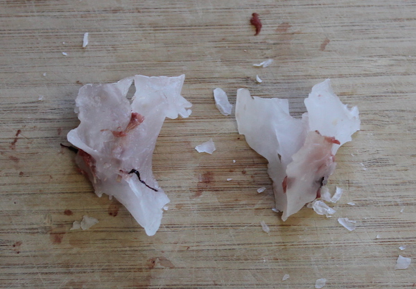
I tried putting the two halves back together and then cutting through the kidney sideways, or transversely. The result is more or less what you would expect:
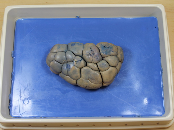
If you ever find a beef kidney to study, you will immediately notice a remarkable difference: They are lumpy instead of smooth. In cows, it seems as if the renal pyramids each grow in their own lobe, instead of merging into a single structure. Why would a lamb kidney be smooth and a beef kidney be lumpy? I don't know, but apparently people can have both kinds. The kidneys of infants are normally “lobulated” like the beef kidney, but then a person's kidneys smooth out as they mature into adults. But every once in a while, the lobes persist into adulthood. I don't think this causes any harm, unless the lobes are mistaken for tumors on an x-ray. An adult with lobulated kidneys is just another example of a “normal anatomic variant.” People sometimes just have different anatomy from each other. Sometimes it causes health problems, sometimes the person never even knows.
Incidentally, I once bought a preserved beef kidney for comparison. I didn't think it was worth the money. One possible benefit of organs from a biology supply company is that the companies can inject colored die into various tubes to help you tell apart the different kinds of tubes. In the kidney, for example, it might be helpful to have color-coding to help distinguish the arteries, the veins, and the urinary passages. However, in general I still recommend fresh organs over preserved organs for most purposes. (And if you are going to opt for injection, definitely buy high-quality specimens from a reputable company. The following photos are of a beef kidney from a discount supplier, and I include them for comparison purposes only.)
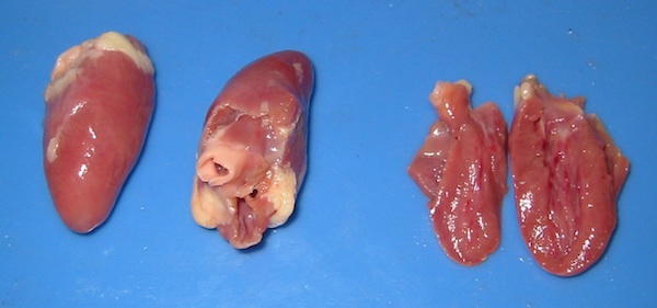
Hearts
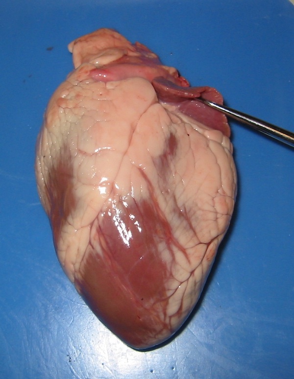
The heart is an oblong muscular organ, which is attached to the lungs and to the two major blood vessels of the body (the aorta and vena cava again) by a large tangle of tubes at the top. There are also two “flaps” at the top, lying alongside the tube tangle. These flaps bear a vague resemblance to the external flaps of human ears, and share their formal name: auricles. The probe in the two pictures below indicates the auricles. At the bottom end, the heart tapers to a muscular peak, called the apex of the heart. Allegedly, this apex bumps against the inside of your sternum when your heart beats, and you feel this in your chest as your heartbeat.
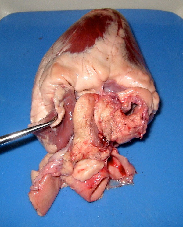
If you insert a probe of some kind into the tubes of the tube tangle, it will probably go all the way inside the heart. You can tell that there are hollow cavities inside the heart, but not all tubes go to the same place. There seems to be more than one cavity inside. To get a better look, you'll need to slice open the heart and look inside.
If you wish to slice through a heart to expose the interior, which way should you slice? If the auricles indicate the “sides” of the heart, then why not slice sideways, through each auricle? That way you can see what is inside underneath both auricles. In the heart shown below, I sliced through one auricle, but I missed the other. The second auricle is hidden behind the top of the right ventricle.
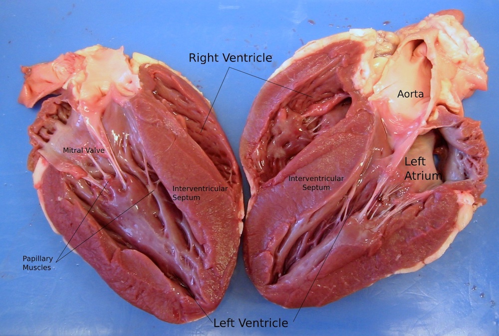
You can see that heart is hollow inside, with two chambers, one chamber underneath each auricle. These are the ventricles. The larger of the two, with thicker more powerful walls, is normally on the left side of the body and is called the left ventricle, and the smaller of the two, with thinner walls, is the right ventricle. (If the heart is made to work by the muscular walls, which of the two chambers do you suppose has the harder job to do?) Within and underneath the auricles are two floppy chambers that look like either the upper portion of the ventricles, or maybe separate chambers above each ventricle. These are the atria. (The right atrium is hidden in the picture above.) A little poking and lifting with a blunt probe (or an old pencil or pen, or a fork for that matter) can reveal tough thin fabric flaps separating the ventricles from the atria. (When these flaps are lying flat against the walls, each atria and ventricle look like a single large chamber.) The visible tendon-like strings apparently hold down the edges of the flap for some reason. If you gently insert the probe through the various tubes in the top of the heart, it will emerge within one of the chambers. All of the tubes on top of the heart open into one of the chambers within.
(The flaps between the atria and ventricles are the mitral valve and the tricuspid valve—one-way valves that let blood flow from the atria to the ventricles, but not vice versa. The tendons—the chordae tendinae, or heart strings—are for holding the valve steady when it is closed so that it doesn't blow inside out like an umbrella in the wind. The little lumpy muscles lining the interior walls that pull on these tendons are the papillary muscles.)
Incidentally, I once read that Leonardo da Vinci, among his many creative explorations of human anatomy, once tried to pour wax into a heart to create casts of the ventricles. I wondered if I could do the same thing, so I bought another heart from the store, along with some paraffin wax. My efforts were rather clumsy, but I think someone else could probably develop a more refined technique, and produce higher-quality casts. (You need to keep the heart warm, to help prevent the melted wax from hardening right away and clogging the openings. But you also don't want to cook the heart. Or maybe you do?)
By the way, do you suppose you could fill a heart with water, then pour it out and measure how much it is? Assuming a lamb's heart is roughly the same size as your own, can you estimate how much blood your heart holds? Can you find your pulse and measure your heart rate, and then estimate how much blood flows through your heart each day? How does this compare to the quantity of food and water you consume each day? What does this tell you about where your blood comes from, and how it moves through your body?
Chicken Hearts
I bought a package of chicken hearts and gizzards from my corner grocery store for about three dollars. On the whole, I don't recommend dissecting chicken hearts. They're too small. They may be interesting as a comparative study, however, especially if you or your students enjoy meticulous work with a magnifying lens.
Livers
Most supermarkets sell sliced beef or calf liver, and I can sometimes find chicken livers in my local grocery store, or whole beef or lamb liver in my nearby Middle Eastern market. However, liver tends to disintegrate with excessive handling, and I haven't found much to observe about supermarket liver anatomically. (This is one case where preserved specimens might be better than fresh. The preservative tends to make the fabric rubbery, so it holds its shape better.) Sometimes you can find large branching tunnels running through a piece of liver. These are presumably portions of the hepatic portal vein, and/or one of the larger branches of it. A liver is basically a sponge with a “tube tree” of veins running through it. (I've managed to find a way to cook sliced liver that makes a dish I actually enjoy eating...except for those veins. Those large veins just won't tenderize.)
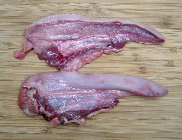
Tongues
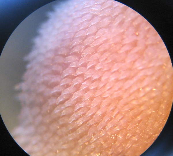
I purchased this lamb tongue from my local Middle Eastern market for about two dollars, and cut it in half with a kitchen shears. Notice the structure—the bulk of the tongue is muscle, surrounded by a covering of skin. Unfortunately, it is hard to identify any more detail than this. I am fairly certain that I found a lingual nerve emerging from the base of the tongue, but it can be hard to tell the difference between nerves and connective fabric.
The lingual papillae at the base of the tongue are large and obvious. The papillae midway down the tongue are smaller, but you can see them fairly easily with the aid of a magnifying lens or low-power dissecting microscope. The following picture is a view of the papillae on the skin from the upper surface of a lamb's tongue, midway between the root and the tip, seen through a microscope.
Stomachs
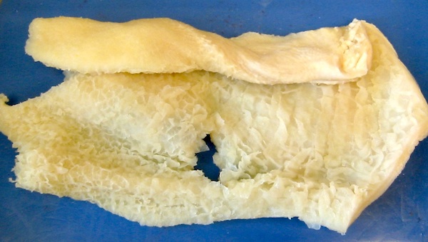
I bought “beef tripe” (i.e. the stomach of a cow) from my grocery store for a few dollars. It consists of a tough leathery fabric — the meaty or muscular stomach wall — covered on the inside by various ripples and folds. (The inner texture will be different depending on which of the cow's several stomachs the tripe comes from.) These folds correspond to the gastric rugae in a human stomach. Why do you suppose the wall of the stomach would be made of muscle? What do you suppose the rugae are for?